Ultrasound-guided carpal tunnel release (CTR) represents a minimally invasive technique gaining traction for treating carpal tunnel syndrome. This approach utilizes
real-time imaging to precisely release the transverse carpal ligament‚ offering potentially quicker recovery and predictable scar healing‚ even bilaterally.
What is Carpal Tunnel Syndrome?
Carpal Tunnel Syndrome (CTS) arises from compression of the median nerve within the carpal tunnel of the wrist. This compression leads to a constellation of symptoms‚ including pain‚ numbness‚ tingling‚ and weakness in the hand and fingers. The condition often stems from repetitive hand motions‚ anatomical factors‚ or underlying health conditions.
Traditional treatment initially focuses on conservative measures like splinting and corticosteroid injections. However‚ when these fail to provide relief‚ surgical intervention—carpal tunnel release—becomes necessary. The goal of CTR is to alleviate pressure on the median nerve by widening the carpal tunnel. Ultrasound guidance offers a modern‚ minimally invasive approach to achieving this release‚ potentially improving outcomes and accelerating recovery compared to traditional methods.
Traditional Carpal Tunnel Release Methods
Historically‚ carpal tunnel release (CTR) has been performed using open surgical techniques. This involves a larger incision in the palm‚ allowing direct visualization and transection of the transverse carpal ligament. While effective‚ open CTR often results in more post-operative pain‚ a longer recovery period‚ and a more prominent scar.
Mini-open CTR represents a modification‚ utilizing a smaller incision. However‚ it still relies on a degree of blunt dissection and doesn’t offer the real-time visualization benefits of newer techniques. Both methods depend on the surgeon’s anatomical knowledge and experience. The emergence of ultrasound-guided CTR provides an alternative‚ aiming to minimize invasiveness and improve recovery‚ challenging the established norms of traditional surgical approaches.
The Rise of Minimally Invasive Techniques
Driven by a desire to reduce patient morbidity and accelerate recovery‚ minimally invasive techniques have gained prominence in carpal tunnel release (CTR). Endoscopic CTR‚ utilizing a small incision and a camera‚ represented an early step towards this goal. However‚ it still requires specialized equipment and a learning curve.
Ultrasound-guided CTR emerges as a further evolution‚ offering a less complex and more accessible minimally invasive option. It avoids the need for a camera‚ relying instead on real-time ultrasound imaging to guide the release. This technique allows for precise ligament transection through a small proximal wrist incision‚ potentially leading to rapid relief and minimal downtime‚ as observed in recent studies.
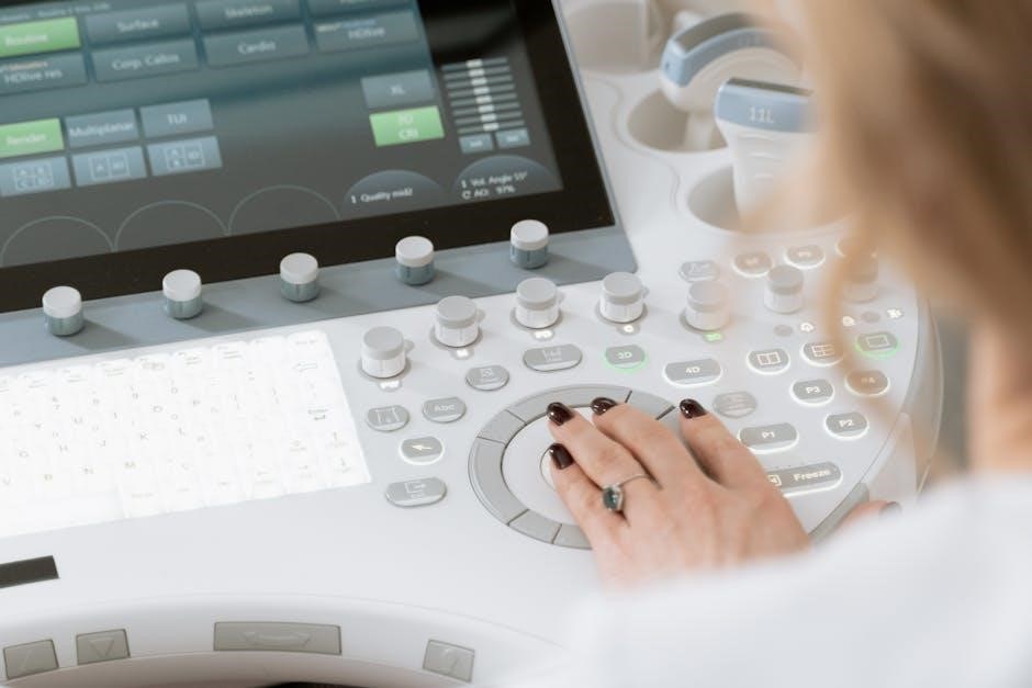
Understanding Ultrasound Guidance in CTR
Ultrasound guidance in carpal tunnel release employs real-time imaging to visualize the transverse carpal ligament and surrounding structures‚ enhancing precision and safety during the procedure.
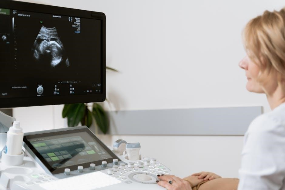
How Ultrasound Imaging Works
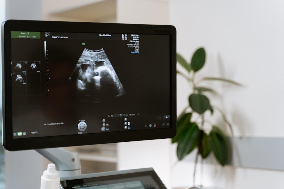
Ultrasound imaging utilizes high-frequency sound waves to create real-time images of structures within the body. A transducer emits these waves‚ which penetrate the tissues and reflect back when they encounter different densities.
These reflected waves‚ or echoes‚ are then processed by a computer to generate a visual representation on a monitor. In the context of carpal tunnel release‚ ultrasound allows surgeons to visualize the transverse carpal ligament‚ the median nerve‚ and other crucial anatomical landmarks.
The technique’s strength lies in its ability to provide dynamic imaging‚ meaning the surgeon can observe movement and adjust the approach accordingly. This is particularly useful when navigating the thread or wire used to transect the ligament‚ ensuring accurate placement and minimizing risk to surrounding neurovascular structures.
Advantages of Real-Time Visualization
Real-time visualization during ultrasound-guided carpal tunnel release (CTR) offers significant advantages over traditional methods. Surgeons can directly observe the transverse carpal ligament and the median nerve‚ enhancing precision and minimizing the risk of injury to critical structures.
This dynamic guidance allows for accurate thread or wire navigation during ligament transection‚ confirming proper positioning before release. The technique facilitates a smaller incision‚ leading to reduced postoperative pain and faster recovery times.
Furthermore‚ real-time imaging enables surgeons to assess the completeness of the release by observing movement of the structures post-transection‚ ensuring adequate decompression of the median nerve.
Limitations of Ultrasound Visualization (Thread Cutting)
Despite its benefits‚ ultrasound visualization during the thread-cutting phase of carpal tunnel release (CTR) presents limitations. Visualizing the cutting thread itself can be challenging due to its small size and the surrounding tissue.
Confirmation of the thread’s precise positioning before ligament transection relies heavily on indirect assessment – observing movement of critical structures when the operator gently tugs on the thread. This method‚ while effective‚ introduces a degree of reliance on tactile feedback and experienced interpretation.
Consequently‚ surgeons must carefully correlate ultrasound imaging with anatomical knowledge to ensure accurate and safe ligament release‚ mitigating potential risks associated with incomplete visualization.
The Procedure: Step-by-Step Breakdown
The ultrasound-guided carpal tunnel release involves patient preparation‚ probe placement for imaging‚ local anesthesia‚ a small incision‚ and precise ligament transection under real-time guidance.
Patient Positioning and Preparation
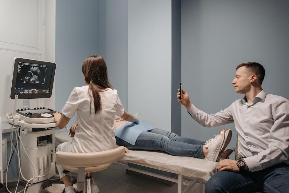
Optimal patient positioning is crucial for successful ultrasound-guided carpal tunnel release. Typically‚ the patient is positioned supine with the affected arm pronated and wrist extended‚ often supported by a gel pad or small roll to achieve a neutral or slightly extended position. This facilitates clear ultrasound visualization of the carpal tunnel structures.
Prior to the procedure‚ the skin over the wrist is meticulously cleaned with an antiseptic solution‚ such as chlorhexidine or iodine‚ to minimize the risk of infection. Sterile drapes are then applied to create a sterile surgical field. The ultrasound probe and associated equipment are also prepared and tested to ensure proper functionality. A time-out procedure is performed to confirm the correct patient‚ site‚ and procedure before commencing.
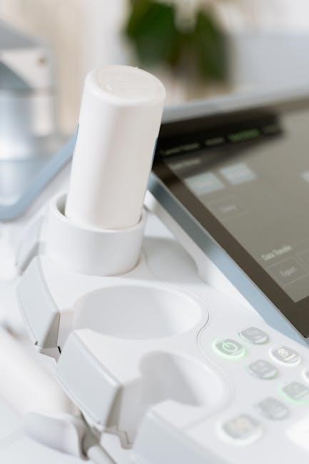
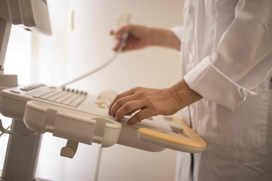
Ultrasound Probe Placement and Imaging
The ultrasound probe‚ typically a high-frequency linear transducer‚ is placed transversely over the volar aspect of the wrist‚ proximal to the carpal tunnel. Gentle pressure is applied to optimize visualization while avoiding compression of the underlying structures. Real-time imaging allows for identification of the median nerve‚ flexor tendons‚ and the transverse carpal ligament.
The operator systematically scans the carpal tunnel to assess the nerve’s size and any potential compression. The goal is to clearly delineate the boundaries of the ligament and confirm its relationship to the median nerve. Precise imaging is essential for guiding subsequent steps‚ ensuring accurate thread/wire insertion and ligament transection.
Local Anesthesia Administration
Following sterile preparation‚ local anesthesia is administered to provide analgesia during the procedure. Typically‚ a combination of lidocaine and bupivacaine is used‚ offering both immediate and prolonged pain relief. Ultrasound guidance can be employed to precisely inject the anesthetic solution around the median nerve and the transverse carpal ligament.
This ensures adequate anesthesia while minimizing the risk of nerve injury. The volume of anesthetic used is carefully controlled to avoid excessive pressure on the nerve. Confirmation of adequate anesthesia is crucial before proceeding with the ligament release‚ often assessed through patient feedback and gentle probing.
Incision and Access
A small‚ proximal wrist incision‚ typically measuring around 1-1.5 cm‚ is created under sterile conditions. This incision provides access for the thread or wire used to transect the transverse carpal ligament. The location is carefully chosen under ultrasound guidance to optimize visualization and minimize risk to surrounding structures.
Dissection is performed through the subcutaneous tissues‚ ensuring careful attention to avoid injury to superficial nerves and vessels. The flexor retinaculum‚ or transverse carpal ligament‚ is then visualized using ultrasound. This minimally invasive approach contrasts with larger‚ more traditional incisions‚ contributing to faster recovery times and reduced scarring.
Technical Aspects of the Release
The procedure involves identifying the transverse carpal ligament‚ inserting a thread/wire under ultrasound guidance‚ and carefully transecting the ligament to achieve release;
Transverse Carpal Ligament Identification
Accurate identification of the transverse carpal ligament (TCL) is paramount for successful ultrasound-guided carpal tunnel release. Utilizing high-resolution ultrasound‚ the surgeon visualizes the TCL as a hyperechoic band spanning the carpal tunnel. This band appears as a bright‚ reflective line on the grayscale image‚ forming the roof of the tunnel.
Careful attention is given to differentiating the TCL from surrounding tendons and structures. Real-time imaging allows for dynamic assessment‚ observing the ligament’s movement during wrist flexion and extension. Precise delineation of the TCL’s boundaries ensures accurate thread/wire insertion and targeted ligament transection‚ minimizing the risk of injury to adjacent neurovascular structures. Proper identification is crucial for a safe and effective release.
Thread/Wire Insertion and Navigation
Following TCL identification‚ a small incision is made‚ and a specialized thread or wire is introduced into the carpal tunnel under continuous ultrasound guidance. The needle’s advancement is carefully monitored to ensure it remains within the tunnel and avoids neurovascular structures. Real-time visualization confirms the thread’s position relative to the TCL.
The thread is then navigated along the course of the ligament‚ often utilizing a gentle sweeping motion. Ultrasound allows the surgeon to track the thread’s progress‚ ensuring complete coverage of the TCL’s span. Precise navigation is essential for effective ligament transection and a successful carpal tunnel release‚ minimizing potential complications.
Ligament Transection Under Ultrasound Guidance
With the thread or wire in position‚ the transverse carpal ligament (TCL) transection begins under direct ultrasound visualization. The operator carefully moves the thread‚ utilizing a sawing or slicing motion to cut the ligament. Real-time imaging is crucial‚ though visualization of the cutting thread itself can be limited.
Confirmation of adequate transection relies heavily on observing movement of the median nerve and other critical structures as the operator gently tugs on the thread. This dynamic assessment‚ guided by ultrasound‚ ensures complete release without causing nerve injury. Precise ligament division is paramount for optimal clinical outcomes.
Confirmation of Release (Structure Movement)
Following presumed ligament transection‚ confirming complete release is vital. Ultrasound assessment focuses on observing dynamic movement of the median nerve‚ flexor tendons‚ and other surrounding structures. The operator gently manipulates the previously inserted thread or wire‚ and assesses for increased gliding and excursion of these structures within the carpal tunnel.
Adequate release is indicated by a noticeable increase in space and freedom of movement. This dynamic confirmation‚ performed under continuous ultrasound guidance‚ compensates for the limited visualization of the cutting thread during the transection process. It ensures a successful outcome and minimizes the risk of incomplete release.
Clinical Outcomes and Efficacy
Studies demonstrate technical and clinical success with ultrasound-guided carpal tunnel release‚ showing positive outcomes in small patient groups with up to six months follow-up.
Reported Success Rates
While comprehensive‚ large-scale data is still emerging‚ initial reports indicate promising success rates for ultrasound-guided carpal tunnel release (CTR). Studies‚ though often involving smaller patient cohorts‚ consistently demonstrate technical feasibility and positive clinical outcomes. Specifically‚ research highlights successful ligament transection and symptom relief in a significant proportion of patients undergoing the procedure.
Comparative analyses against traditional open and mini-open techniques are beginning to show comparable‚ and in some cases‚ improved results. The ability to visualize structures in real-time contributes to accurate ligament release‚ potentially minimizing complications. However‚ it’s crucial to acknowledge that success rates can vary based on surgeon experience and patient-specific factors. Further investigation through robust clinical trials is ongoing to establish definitive benchmarks.
Comparison with Open and Mini-Open Techniques
Ultrasound-guided carpal tunnel release (CTR) is increasingly evaluated alongside established open and mini-open surgical approaches. Initial meta-analyses and comparative studies suggest that ultrasound guidance offers comparable efficacy in relieving carpal tunnel syndrome symptoms. A key advantage lies in its minimally invasive nature‚ resulting in smaller incisions and potentially reduced postoperative pain.
While traditional methods provide a direct surgical field‚ ultrasound guidance allows for real-time visualization of critical structures‚ potentially enhancing precision. Recovery times are often reported as faster with the ultrasound technique‚ contributing to quicker return to function. However‚ long-term outcomes require further investigation to definitively determine superiority across all patient profiles.
Long-Term Clinical Outcomes (6-Month Follow-up)
Studies examining the 6-month follow-up period after ultrasound-guided carpal tunnel release (CTR) demonstrate promising clinical success. Reported outcomes indicate significant improvements in symptom scores‚ grip strength‚ and nerve conduction studies in a majority of patients. Technical success rates are also high‚ with minimal reported complications during this timeframe.
However‚ it’s crucial to note that most published data currently stems from smaller patient samples. While these initial results are encouraging‚ larger‚ multi-center trials are needed to validate these findings and establish long-term durability. Continued monitoring is essential to assess sustained symptom relief and potential late complications.
Recovery and Rehabilitation
Post-operative pain is typically managed with standard analgesics‚ and the recovery timeline is notably quick with ultrasound-guided CTR‚ allowing for early mobilization.
Post-Operative Pain Management
Following ultrasound-guided carpal tunnel release‚ effective pain management is crucial for a comfortable recovery; Typically‚ a combination of over-the-counter analgesics‚ such as acetaminophen or ibuprofen‚ proves sufficient for controlling discomfort. In some cases‚ a short course of prescription pain medication may be considered‚ particularly in the initial 24-48 hours post-procedure.
Patients are generally advised to apply ice packs to the operative site for the first few days to minimize swelling and alleviate pain. Elevation of the hand can also contribute to reducing edema. It’s important to follow the surgeon’s specific instructions regarding medication dosage and frequency. Most individuals experience a gradual decrease in pain within the first week‚ transitioning to mild discomfort that is easily managed with non-pharmacological methods.
Expected Recovery Timeline
The recovery following ultrasound-guided carpal tunnel release is generally swift‚ a key advantage of this minimally invasive technique. Patients often experience immediate improvement in symptoms‚ with significant pain reduction within days. Light activities can usually be resumed within a week‚ gradually increasing as tolerated.
Full grip strength typically returns within 4-6 weeks‚ though some residual tenderness or mild discomfort may persist for up to three months. The predictable and quick recovery allows for consideration of bilateral releases on the same day‚ a practice less common with traditional methods. Most individuals return to all normal activities‚ including work and hobbies‚ within 6-8 weeks post-procedure.
Bilateral Release Feasibility & Scar Healing
A significant benefit of ultrasound-guided carpal tunnel release is the increased feasibility of performing bilateral procedures simultaneously. Historically‚ surgeons often avoided releasing both hands on the same day due to unpredictable palmar scar healing‚ potentially hindering function.
However‚ the minimally invasive nature of the ultrasound technique results in a remarkably predictable and rapid recovery. This allows patients to have both carpal tunnels addressed in a single session‚ minimizing overall downtime and accelerating return to full activity. Scarring is typically minimal‚ often appearing as a small‚ inconspicuous mark‚ further contributing to a positive patient experience.
Potential Risks and Complications
While generally safe‚ ultrasound-guided carpal tunnel release carries potential risks including nerve injury‚ infection‚ and incomplete ligament release‚ requiring careful technique.
Nerve Injury
Nerve injury represents a significant‚ though relatively uncommon‚ complication associated with any carpal tunnel release procedure‚ including the ultrasound-guided technique. The median and ulnar nerves are in close proximity to the surgical field‚ making them vulnerable to direct trauma during ligament transection.
Careful attention to ultrasound imaging is crucial to visualize nerve location and avoid inadvertent injury. Experienced surgeons meticulously monitor the cutting thread’s position relative to these structures. Post-operative symptoms such as numbness‚ tingling‚ or weakness warrant immediate evaluation to rule out nerve compression or damage. Complete nerve injury is rare‚ but transient paresthesias are more frequently reported and typically resolve with time.
Infection
While ultrasound-guided carpal tunnel release (CTR) is a minimally invasive procedure‚ the risk of infection‚ though low‚ remains a potential complication. As with any surgical intervention‚ bacteria can enter the incision site‚ leading to localized or systemic infection. Strict adherence to sterile technique during the procedure is paramount in minimizing this risk.
Post-operative wound care instructions‚ including keeping the incision clean and dry‚ are crucial for patients. Signs of infection – increasing pain‚ redness‚ swelling‚ pus drainage‚ or fever – should prompt immediate medical attention. Prompt diagnosis and treatment with antibiotics are typically effective in resolving infections.
Incomplete Release
An incomplete release of the transverse carpal ligament represents a potential complication in ultrasound-guided carpal tunnel release (CTR). This occurs when the ligament isn’t fully transected‚ continuing to compress the median nerve and resulting in persistent symptoms. Accurate identification and complete transection of the ligament under real-time ultrasound guidance are vital to avoid this outcome.
Confirmation of adequate release‚ assessed by observing movement of the critical structures during thread manipulation‚ is crucial during the procedure. If symptoms persist post-operatively‚ further evaluation‚ potentially including repeat ultrasound imaging‚ may be necessary to determine if a revision surgery is required to complete the ligament release;
Patient Selection Criteria
Ideal candidates exhibit typical carpal tunnel syndrome symptoms‚ while contraindications include bleeding disorders or infections. Pre-operative evaluation confirms diagnosis and assesses suitability.
Ideal Candidates for Ultrasound-Guided CTR
Patients experiencing symptoms consistent with carpal tunnel syndrome‚ such as nocturnal paresthesia‚ numbness‚ tingling‚ and weakness in the hand‚ are generally considered ideal candidates. Diagnostic confirmation through nerve conduction studies is often beneficial. Individuals seeking a minimally invasive approach with a potentially faster recovery timeline may particularly benefit from this technique.
Those who have failed conservative treatments – including splinting‚ activity modification‚ and corticosteroid injections – are also strong candidates. Furthermore‚ patients desiring a return to quick hand function‚ and those potentially suitable for bilateral simultaneous release‚ due to the predictable recovery‚ are well-suited for ultrasound guidance. The absence of significant comorbidities impacting healing is also a positive factor.
Contraindications
While ultrasound-guided carpal tunnel release (CTR) is generally safe‚ certain conditions present contraindications; Active infection in the operative hand or systemic infection represents a clear contraindication‚ as does significant coagulopathy or bleeding disorder. Patients on full anticoagulation may require temporary cessation of medication‚ guided by their physician.
Severe‚ uncontrolled diabetes can impair wound healing and increase infection risk‚ making these patients less suitable. Pre-existing skin conditions at the surgical site‚ such as severe dermatitis‚ could also complicate the procedure. Finally‚ patients with unrealistic expectations regarding surgical outcomes or those unable to follow post-operative instructions should be carefully evaluated.
Pre-Operative Evaluation
A thorough pre-operative evaluation is crucial for successful ultrasound-guided carpal tunnel release (CTR). This begins with a detailed medical history‚ focusing on comorbidities like diabetes and bleeding disorders; A comprehensive neurological examination assesses symptom severity‚ nerve function‚ and identifies potential contributing factors. Nerve conduction studies (NCS) and electromyography (EMG) confirm the diagnosis and severity of carpal tunnel syndrome.
Patients should be assessed for anatomical variations that might influence the procedure. Discussion of risks‚ benefits‚ and alternatives is essential‚ ensuring informed consent. Finally‚ evaluation of hand dominance and activity level helps tailor post-operative rehabilitation plans.
Future Directions and Research
Ongoing studies explore technological advancements in ultrasound guidance for CTR‚ aiming to improve visualization and precision. Expanding applications and long-term efficacy are key research areas.
Ongoing Studies and Clinical Trials
Currently‚ several investigations are focused on solidifying the role of ultrasound-guided carpal tunnel release (CTR) as a standard treatment option. These studies aim to gather more robust data regarding its technical success and long-term clinical outcomes‚ particularly beyond the six-month follow-up period commonly reported. Researchers are actively comparing the ultrasound-guided technique against traditional open and mini-open surgical approaches‚ utilizing randomized controlled trials to minimize bias and ensure reliable results.
A significant area of focus involves evaluating the consistency of nerve injury rates and assessing the effectiveness of various rehabilitation protocols post-operatively. Furthermore‚ clinical trials are investigating the potential benefits of utilizing advanced ultrasound technologies to enhance visualization of the cutting thread and confirm accurate ligament transection‚ addressing current limitations in the procedure.
Technological Advancements in Ultrasound Guidance
Innovations in ultrasound technology are actively addressing the limitations currently experienced with ultrasound-guided carpal tunnel release (CTR). Researchers are exploring higher-resolution imaging systems to improve visualization of the transverse carpal ligament and surrounding structures‚ particularly the cutting thread during transection. This enhanced clarity aims to reduce the reliance on operator feel and improve precision.
Furthermore‚ advancements in image processing software are being investigated to provide real-time feedback on ligament release‚ potentially confirming complete transection through dynamic assessment of structure movement. The integration of elastography‚ a technique that measures tissue stiffness‚ could also offer valuable information regarding ligament integrity. These developments promise to refine the technique and enhance its overall efficacy and safety.
Expanding Applications of the Technique
Beyond standard carpal tunnel syndrome‚ exploration is underway to assess the utility of ultrasound-guided release in variations of the condition. This includes cases with anatomical variations or those presenting with atypical symptom patterns. The minimally invasive nature of the procedure lends itself well to revision surgeries following previous failed carpal tunnel releases‚ offering a less disruptive approach.
Researchers are also investigating the potential for applying ultrasound guidance to other entrapment neuropathies affecting the upper extremity. The precision offered by real-time imaging could prove beneficial in releasing other ligaments or structures compressing peripheral nerves. Ultimately‚ the goal is to broaden the scope of ultrasound-guided techniques to address a wider range of nerve compression syndromes.
No Responses